Environmental DNA in subterranean biology: range extension and taxonomic implications for Proteus
Povezani članci
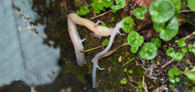
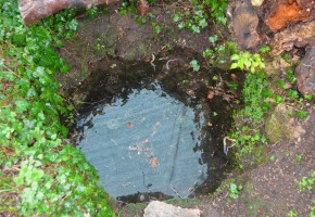
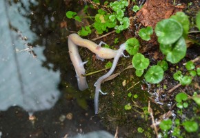
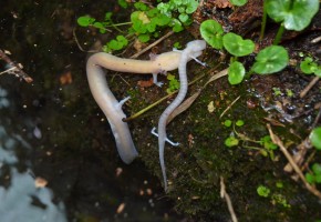
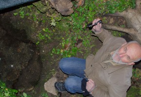
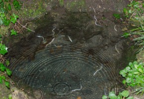
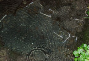
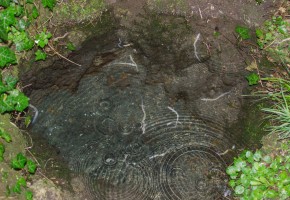
Photo: Zlatko Grizelj
Špela Gorički, David Stanković, Aleš Snoj, Matjaž Kuntner, William R. Jeffery, Peter Trontelj, Miloš Pavićević, Zlatko Grizelj, Magdalena Năpăruş-Aljančič & Gregor Aljančič
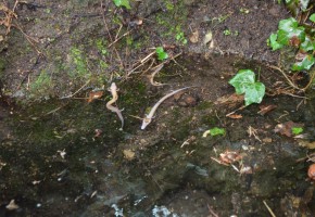
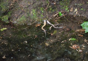
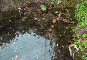
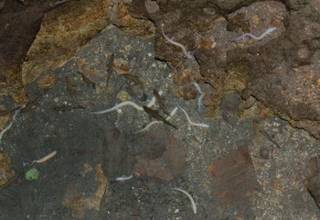
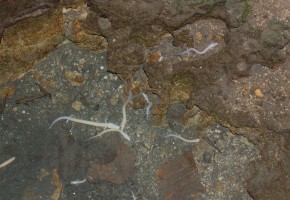
Photos: Zlatko Grizelj
Abstract
Europe’s obligate cave-dwelling amphibian Proteus anguinus inhabits subterranean waters of the north-western Balkan Peninsula. Because only fragments of its habitat are accessible to humans, this endangered salamander’s exact distribution has been difficult to establish. Here we introduce a quantitative real time polymerase chain reaction-based environmental DNA (eDNA) approach to detect the presence of Proteususing water samples collected from karst springs, wells or caves. In a survey conducted along the southern limit of its known range, we established a likely presence of Proteus at seven new sites, extending its range to Montenegro. Next, using specific molecular probes to discriminate the rare black morph of Proteus from the closely related white morph, we detected its eDNA at five new sites, thus more than doubling the known number of sites. In one of these we found both black and white Proteus eDNA together. This finding suggests that the two morphs may live in contact with each other in the same body of groundwater and that they may be reproductively isolated species. Our results show that the eDNA approach is suitable and efficient in addressing questions in biogeography, evolution, taxonomy and conservation of the cryptic subterranean fauna.
Introduction
The olm, Proteus anguinus Laurenti 1768, is a large amphibian endemic to subterranean waters of the Dinaric Karst, with a known range stretching between north-eastern Italy and southern Bosnia and Herzegovina. Despite the relatively broad expanse (60,000 km2) of karst topography in this region and decades of field surveys, Proteus has only been documented at around 300 sites1,2,3,4. These sites include caves where Proteus is recorded by visual observation or trapping, and springs where specimens may emerge during seasonal flooding5. While groundwater pollution and destruction of subterranean habitat are obvious threats to Proteus6,7, the negative anthropogenic impact cannot be determined without a reliable methodology to establish and monitor its presence.
Even with advances in speleobiology, progress in defining the true geographic distribution and diversity of its populations has been slow8. For example, despite much speculation, no physical evidence of the presence of Proteus in the Dinaric Karst of Montenegro has been documented. Furthermore, as recently as in 1986, a unique, darkly pigmented non-troglomorphic population of Proteus was discovered in south-eastern Slovenia9 and described as the subspecies Proteus anguinus parkelj10. The results of subsequent morphometric analyses11,12,13 supported its distinct taxonomic status, and phylogeographic analyses of mitochondrial DNA (mtDNA)14,15 confirmed the distinctiveness of its lineage, one, however, that is deeply nested in the Proteus phylogeny. The population of black Proteus has been documented at only four sites in an area of less than 2 km2 (refs 1, 7 and 16). In the same geographic region, but presumably in a different hydrogeological formation17,18, a closely related lineage14 of the troglomorphic, white Proteus subspecies (Proteus anguinus anguinus) has also been recorded at nine sites1,9 (also A. Hudoklin, pers. comm. 20 July 2015). If two such morphs co-existed in the same local habitat without hybridizing, they would likely be reproductively isolated from each other by an intrinsic barrier. However, this simple and powerful test of species status is rarely available in obligate subterranean organisms, because their habitat is patchy and their populations are usually fragmented and physically strongly isolated from each other19,20,21.
To detect species like Proteus, that are rare and difficult to observe with classical methods, detection of specific DNA released into the environment (environmental DNA or eDNA) is particularly useful22,23,24,25,26,27. The ubiquitous nature of DNA in aquatic environments and its rapid diffusion from its source means that in theory the presence of a specific animal can be detected anywhere within the water body and not just at its point of origin28,29. Furthermore, as DNA released into most environments becomes quickly degraded, the eDNA approach detects the recent presence of target species30 without the need for direct observation or trapping.
In this study we used Proteus individuals from the laboratory to develop a set of eDNA detection assays based on quantitative real-time polymerase chain reaction (qPCR) to examine the presence of Proteus in karst aquifers, where physical detection is difficult or impossible. First, we developed a SYBR green (Applied Biosystems) qPCR assay to search for Proteus eDNA in spring and cave water samples from the under-explored southernmost edge of the known range of Proteus in Herzegovina (southern Bosnia and Herzegovina) and outside of it in Montenegro. Second, we developed a TaqMan (Applied Biosystems) qPCR assay to discriminate the black Proteus eDNA from the white Proteus eDNA. Using this assay, we conducted a systematic inventory of Proteus in Bela Krajina (south-eastern Slovenia) to verify selected historic records of the white Proteus, to determine the maximum range of the black Proteus and to test for possible co-occurrence of the two morphs.
Results
Proteus eDNA detection by qPCR
The sample validation procedure for the observed outcomes of qPCR tests is illustrated in Fig. 1. The Supplement lists the lower limit of detection and the confirmation of assay specificity for both SYBR and TaqMan qPCR assays. No false positives were observed.
Evaluation of Proteus eDNA presence in a sample from the observed outcome of (a) SYBR qPCR assay and (b) TaqMan qPCR assay. Two mitochondrial DNA (mtDNA) regions (“genes”) were searched for in (a) and one mtDNA region was searched for in (b). The first run in both (a) and (b) included two concentrations (“dilutions”) of the template (data were pooled in b). All assays were performed in three parallel reactions (“wells”). See also the Methods section.
Out of 23 sites in Herzegovina examined for eDNA by the SYBR qPCR assay, four were verified to harbour Proteus (confirmed through visual encounter by a reliable informant). Out of these, two scored positive and one plausible for its eDNA while the fourth was negative. In Bela Krajina (Slovenia), only one verified site was included in the analyses by the TaqMan qPCR assay and it scored positive. Out of additional four likely sites (within the known range of the black Proteus), three also were positive and one was negative.
In our estimations during sampling, the flow of the springs where Proteus eDNA was detected varied from springs and wells with discharge rates as low as 0.1 L/s to as high as 2000 L/s (see Supplementary Table S1). The maximum water temperature recorded during sampling was an exceptional 17 °C, with the median at 11.7 °C (see Supplementary Table S1). Once sampled, DNA in water degraded within several days when stored at 4 °C. By contrast, storage of dry filters at −20 °C sufficiently preserved the integrity of the DNA for at least two months, and storage of isolated DNA at −20 °C for at least four months.
Detection of Proteus by eDNA in Herzegovina and Montenegro
In Herzegovina (Fig. 2), the springs Londža, Muša (no. 1 in Supplementary Table S1), Nezdravica (no. 14) and Izvor Bregave (no. 20) showed weak signals (one positive signal in three replicates) representing only one of the genes and were negative in the re-run. Hence, these samples are categorised as uncertain for the presence of Proteus eDNA (possibly at the limit of detection, although we cannot completely exclude contamination despite the precaution mechanisms). A private well in Gornji Trebižat (no. 2) and Vrelo Vakuf (no. 4) showed a weak signal (one positive signal in three replicates) representing only the mitochondrial control region, and this signal (one positive signal in three replicates) was again observed in the re-run. Therefore, these samples are categorised as plausible for containing Proteus eDNA. Here contamination is less likely as the signal was observed in independent runs. Perića Mlin (no. 3) and Kajtazovo Vrelo (no. 5) were positive for one gene, whereas the samples from Bunar kod Kuće Mehe Dizdarevića (no. 10) and Česma izpod Pogledovače (no. 11) were positive for both genes. Thus, these localities are interpreted as positive for the presence of Proteus eDNA.
Categorisation of sites is explained in Fig. 1a and in the Methods section. The map was created using ArcGIS Desktop 10.3.1 (Esri 2015). Basemap used: World Street Map (Esri 2015). Source of hydrogeological data: http://diktas.iwlearn.org/im/hydrogeological-map-of-the-dinaric-karst (last accessed 5 October 2016).
In Montenegro (Fig. 3), the cave Sopot (no. 24 in Supplementary Table S1) and the spring Izvor Grahovo 1 (no. 29) showed weak signals (one positive signal in three replicates) representing only one of the genes and were negative in the re-run. Therefore, these signals are interpreted as uncertain for the presence of Proteus eDNA. The spring Šanik (no. 33) also showed a weak signal representing only the 16S rRNA gene (one positive out of three replicates), but in this case the same signal was observed again in three separate re-runs. Therefore, this sample is interpreted as plausible to contain Proteus eDNA, and here contamination is less likely as the signal was observed in four independent runs.
Categorisation of sites is explained in Fig. 1a and in the Methods section. The map was created using ArcGIS Desktop 10.3.1 (Esri 2015). Basemap used: World Street Map (Esri 2015). Source of hydrogeological data: http://diktas.iwlearn.org/im/hydrogeological-map-of-the-dinaric-karst (last accessed 5 October 2016).
Detection of black and white Proteus by eDNA in Bela Krajina (Slovenia)
Proteus eDNA was confirmed in six out of 19 samples analysed from Bela Krajina (Fig. 4). Except for Otovski Breg (no. 45 in Supplementary Table S1), a verified site with white Proteus, all of the positive sites were springs along the Dobličica River. Following the direction of the Dobličica River flow, there was a gradient of relative concentration of Proteus eDNA in these springs, as deduced from cycle threshold (Ct) values in combination with the fraction of positive replicates in each sample. In the south-to-north and west-to-east directions, these fractions were as follows: Izvir ob Dobličici BK D3 (no. 51) 6/6, Izvir ob Dobličici BK D4 (no. 52) 4/6, Izvir ob Izlivu Jelševnice v Dobličico BK A2 (no. 47) and Šprajcarjev Zdenec (no. 41) 6/6, Izvir v Svibniku (no. 54) 2/6 + 2/3, Planinec (no. 55) 1/6 + 0/3. Outside the immediate area of the Dobličica River, the Otovski Breg (no. 45) sample was positive in 5/6 reactions, and Izvir Obrščice (no. 40) in only 1/6 + 0/3 reactions. The latter and Planinec were not analysed further as the presence of ProteuseDNA in these springs was uncertain. All other analysed samples were negative for the presence of Proteus eDNA. Following our analyses, on the evening of November 1, 2016, a young black Proteus was observed by the last two co-authors in Planinec.
Circles and squares depict the results of eDNA analyses; stars and triangles represent known localities of the black or the white Proteus, respectively. Categorisation of sites is explained in Fig. 1b and in the Methods section. Doblička Gora and Poljanska Gora are the south-eastern foothills of the Kočevski Rog Plateau. The map was created using ArcGIS Desktop 10.3.1 (Esri 2015). Source of basemap: digital elevation model at 1:10,000 (http://www.e-prostor.gov.si/si/zbirke_prostorskih_podatkov/topografski_in_kartografski_podatki/digitalni_model_visin/digitalni_model_visin_5_x_5_m_dmv_5/, last accessed 5 October 2016).
Once Proteus eDNA was confirmed in a sample, it was tested further to determine if the eDNA belonged to the black or the white Proteusmorph (Fig. 5). We thus detected black Proteus eDNA in five samples, all taken in springs along the Dobličica River, while the Otovski Breg (no. 45) sample was negative for black Proteus eDNA (0/6 reactions). The fraction of positive reactions again followed a northward and eastward gradient: Izvir ob Dobličici BK D3 (no. 51) 6/6, Izvir ob Dobličici BK D4 (no. 52) 3/6, Izvir ob Izlivu Jelševnice v Dobličico BK A2 (no. 47) 6/6, Šprajcarjev Zdenec (no. 41) 3/6, Izvir v Svibniku (no. 54) 2/6. We next analysed the same six samples for white Proteus eDNA. As expected, white Proteus eDNA was confirmed in the Otovski Breg sample (no. 45; 3/6), a known white Proteus site. Importantly, however, white ProteuseDNA was found in Šprajcarjev Zdenec (no. 41; 2/6 + 3/3), which was also positive for black Proteus eDNA. All other samples that were positive for black Proteus eDNA were negative for white Proteus eDNA. Sequencing of the PCR products amplified in the sample from Šprajcarjev Zdenec confirmed that both the black and the white haplotype were present in the sample.
(a) Distribution of eDNA specific for (b) black and (c) white Proteus in spring samples in Bela Krajina (Slovenia). Circles and squares depict the results of eDNA analyses; stars and triangles represent known localities of the black or the white Proteus, respectively. The map was created using ArcGIS Desktop 10.3.1 (Esri 2015). Source of basemap: digital elevation model at 1:10,000 (http://www.e-prostor.gov.si/si/zbirke_prostorskih_podatkov/topografski_in_kartografski_podatki/digitalni_model_visin/digitalni_model_visin_5_x_5_m_dmv_5/, last accessed 5 October 2016). Sources of hydrogeological and geological layers: refs 17, 18 and 51.
Discussion
Below we evaluate the basic parameters of our eDNA assay and its effectiveness in detecting in the laboratory as well as in different subterranean habitats.
A suitable eDNA assay for Proteus must be able to detect trace amounts of highly diluted DNA released by potentially very small populations. The qPCR technique is commonly used in achieving this goal22,31. When compared to classical PCR in combination with cloning and sequencing27, the real-time qPCR approach significantly improves the efficiency of eDNA detection and reduces the possibility of contamination as post-PCR analysis is omitted. Our tests showed that this method is suitable for detection of Proteus eDNA, including under the conditions encountered at karst springs.
A compromise between at least three factors is necessary for an optimal use of the eDNA assay for Proteus: (1) the time of sampling, (2) the amount of water filtered and (3) the lower detection limit of the method. The discharge from karst springs typically varies significantly through seasons in response to precipitation in the catchment area, with many springs inactive during the dry season32,33. As a consequence, sampling when water levels are optimal may be a challenge. When water levels are very high, eDNA may become too diluted or dispersed for detection. The latter may have been the case in a few springs in Herzegovina, which therefore required a repeated sampling to detect the presence of Proteus eDNA. Another concern when sampling for Proteus eDNA is collecting water samples without disturbing the sediment at very low water levels, while higher water levels may increase sediment transport through the karst aquifer. Even though sediment potentially holds more eDNA34,35, it also prevents efficient filtration. Furthermore, sediment can be a source of PCR inhibitors36,37. Monitoring PCR inhibition with the addition of synthetic DNA and corresponding primers and probes to each reaction, and diluting template DNA when inhibition is detected, is essential when analysing environmental samples for eDNA.
Exposed DNA in water gradually decays predominantly due to the effects of UV-light, heat and decomposition by microorganisms31,38,39. Because of the absence of light and due to the relatively low and constant temperatures of groundwater inhabited by Proteus(Supplementary Table S1; see also ref. 1), we expect eDNA to be relatively stable, which presumably facilitates detection. Furthermore, the eDNA fragments targeted in our assays are 100–150 bp long, i.e. short enough to persist in the environment40 despite degradation processes. As factors that affect DNA degradation may be less detrimental in subterranean streams, eDNA transport distance is likely increased in this habitat. Since eDNA transport in streams is greatly affected by discharge rates41,42, we timed the sampling, whenever and wherever it was possible, to the lowest water level and discharge rates that still allowed for efficient sampling.
The mitochondrial control region and flanking DNA and the 16S rRNA gene were chosen for our eDNA assay because a large set of sequences in Proteus is available for comparison and primer/probe design (see Supplementary Information). We believe this approach minimises the risk of not detecting a Proteus population due to an unknown variation in sequence. This especially applies to the primer set designed to bind to the conserved 16S rRNA gene, which appears to be general enough to be used in detection of any population within as well as outside the known range of Proteus. The primer pair designed to amplify a fragment of the control region, on the other hand, targets a more variable region of the mitochondrial genome. Therefore, the possibility that it could not bind efficiently to the hypothetical Montenegro population, which may be genetically distinct from all other known populations, cannot be excluded.
Compared to classical approaches of visual surveying or trapping, the eDNA analysis substantially improves our ability to detect Proteus in groundwater. This is most clearly observable in the results of the Bela Krajina survey, where the number of known sites with the black Proteusmore than doubled after a single sampling. Under optimal water-level conditions, the sampling protocol developed here for Proteus eDNA is expected to yield at least a 75% detection rate at densities of at least one animal per 256 m3 of water (see Supplementary Information). Although the number of sites used to validate the detection probability and accuracy of the method was low, laboratory tests (see Supplementary Information) and hydrogeological data strongly support the results obtained for springs with hitherto unknown status. Similar detection probabilities were reported for epigean aquatic vertebrate species (e.g. refs 25, 42 and 43).
Testing the usefulness of the eDNA assay in field research in Herzegovina resulted in the detection of Proteus at locations where it has not been previously recorded. In the most recent list of localities in Bosnia and Herzegovina3, only seven out of a total of 57 are known in the greater Trebižat River area. We discovered four new Proteuslocalities by the eDNA method and two very likely to harbour Proteus. Following our analyses, the presence of Proteus was visually confirmed by cave diving at the spring Kajtazovo Vrelo (no. 5 in Supplementary Table S1; Z. Vlaho, pers. comm. 29 June 2016).
Here we also report the evidence for the existence of Proteus in Montenegro, based on a plausible sample of Proteus eDNA recorded at the spring Šanik (no. 33 in in Supplementary Table S1). Šanik is located 1.7 km from the nearest Proteus locality in Herzegovina (B. Lewarne, pers. comm. 22 June 2014). The proximity of the two sites suggests that they may be hydrogeologically connected and therefore may share a common Proteus population. Alternatively, they may depend on the same catchment area, which could include an upstream Proteus locality potentially serving only as a donor site for eDNA influx into Šanik. Since no hydrogeological surveys have been conducted in the region, neither a connection of Šanik to the locality in Herzegovina nor the location of its catchment area has been determined. Nonetheless, preliminary results suggest that the two cave systems could be hydrogeologically isolated from each other (B. Lewarne, pers. comm. 2 October 2016). Our results therefore favour the possibility that Proteus individuals are actually present at the sampled location and that the southern range of Proteus extends into the Dinaric Karst of Montenegro.
Next, using the eDNA assay, we report the discovery of new sites that may harbour the rare black Proteus, while its presence was visually confirmed in yet another one, which showed a faint trace of ProteuseDNA. Three of these springs lie outside the limits of its known range and represent an extension of its presumed range north-eastward, along with the general direction of the flow of the Dobličica River. The distance of the new easternmost site, the spring Planinec (no. 55 in Supplementary Table S1), from the nearest previously visually confirmed site, Kanižarica16, is 1.2 km (the actual extent of the cave system is unknown). Since we confirmed by visual observation that the occurrence of Proteus eDNA in a spring is indicative of its actual presence in close proximity, the present knowledge confines the black Proteus between the high plateau Kočevski Rog – which is lacking surface streams – in the west, along the Dobličica River to the confluence with the stream Pački Potok in the east.
As in most aquatic cave animals, ranges of Proteus populations are probably historically determined and highly restricted by the boundaries of present-day subterranean hydrogeological networks15. The white Proteus population appears to occupy a larger range, including sites in the Dolenjska region west of Kočevski Rog14. It should be noted, however, that while the maximum span of the black Proteusrange to the east, north and south was established in the present study, the extent of its distribution to the west remains unknown. The observed descending gradient in concentration of eDNA following the Dobličica main stream flow appears to reflect a complex local network of underground connections that could receive inflow through both southwest-to-northeast and northwest-to southeast oriented faults as proven also by the water tracings made in the region17,18 (see Fig. 5). Therefore, the core of the black Proteus’ distribution probably includes the contact zone between the Kočevski Rog Plateau and the Bela Krajina Plain as well as the south-eastern parts of Kočevski Rog. This conclusion is consistent with earlier predictions, when only one9 or two sites10 were known.
The discovery of the eDNA of both black and white Proteus syntopically in the spring Šprajcarjev Zdenec (no. 41 in Supplementary Table S1) represents direct evidence suggesting that these two populations may be in contact with each other. The finding is strongly supported by an existing (intermittent) hydrogeological connection17 with Otovski Breg, a nearby site occupied by the white Proteus (no. 45 in Supplementary Table S1). Because the relative concentration of eDNA of the white Proteus in Šprajcarjev Zdenec was similar to the relative concentration detected at Otovski Breg (sampled at around the same date), which cannot be explained by the difference in discharge rates of the two springs alone, we believe that it reflects the actual presence of the white Proteus in Šprajcarjev Zdenec. Passive eDNA dispersal would likely result in the presence of white Proteus eDNA in the spring closest to Šprajcarjev Zdenec (Izvir v Svibniku; no. 54 in Supplementary Table S1), irrespective of a later sampling date and alongside black ProteuseDNA found there. As our analysis showed the relative concentration of white Proteus eDNA in Šprajcarjev Zdenec to be similar to the relative concentration of black Proteus eDNA in this same spring, but at the same time we failed to detect white Proteus eDNA in nearby Izvir v Svibniku, we expect repeated additional sampling would support this conclusion.
Rare cases of subterranean syntopic occurrence of closely related lineages are valuable for the study of the poorly understood mechanisms of speciation and differentiation within the subterranean realm (e.g. ref. 44). The distribution of the black and white Proteus eDNA in Bela Krajina is in agreement with the existence of a potential reproductive barrier between these two lineages, at least regarding female mating preferences. Assuming gene flow between the two lineages, substantial introgression of the white Proteus mtDNA would predict the presence of white Proteus eDNA in the springs to the west of Šprajcarjev Zdenec, which was not detected. Similarly, if inter-lineage mating regularly occurred in the other direction, we would expect black Proteus eDNA to appear in the sample at Otovski Breg, a site which was found to harbour only white Proteus eDNA. Significantly, despite a low degree of sequence divergence between the two populations observed in the mitochondrial control region14,15,45, none of the comparative studies to date have detected any signs of their interbreeding, e.g. haplotype sharing14,15 or intermediate morphology10,12,13,46,47,48. In combination with these observations, eDNA data suggest that the two populations may represent independent species, but additional analyses are needed to resolve the taxonomic status of the present as well as of other apparently monophyletic groups of Proteus.
In conclusion, we have demonstrated that the qPCR-based eDNA method can be utilised for a rapid detection of a rare subterranean species inhabiting karst groundwater. Due to its high sensitivity (see Supplementary Information) and general applicability, the SYBR qPCR-based eDNA assay is appropriate for large-scale inventories of Proteusin groundwater throughout the Dinaric Karst, while the high specificity of the TaqMan qPCR-based eDNA assay makes this approach suitable for monitoring the distribution of closely related populations or taxa. Furthermore, as suggested by the results of the Bela Krajina survey in particular, the qPCR-based approach enables us to assess the relative abundance of Proteus eDNA in groundwater over a small spatial scale. Finally, we have shown that the eDNA approach can also be helpful in identifying potentially sympatric populations in the cryptic subterranean environment and therefore can be useful in the study of evolutionary history and taxonomy of subterranean taxa.
Methods
Study Design
Development of our methodology to detect traces of Proteus eDNA in water involved the following steps (see Supplementary Information): (1) development of specific oligonucleotides for eDNA detection with qPCR, (2) testing the specificity of the oligonucleotides on tissue samples, (3) testing the lower detection limit of the method in laboratory conditions and (4) testing the performance of the method in nature, at three verified sites in Slovenia (SYBR qPCR only). Two broad geographic regions in the south-eastern part of the Dinaric Karst were then investigated for the presence and distribution of Proteus using the SYBR qPCR assay, while the distribution of two morphs of Proteus in south-eastern Slovenia was surveyed using the TaqMan qPCR assay. Our eDNA methodology is in line with general recommendations for eDNA sampling, analysis and reporting49.
Water filtration and eDNA extraction
At field sampling sites, 10 (exceptionally) to 20 L of water were collected taking care not to disturb the sediment during sampling. Samples were collected in brand-new 5- or 10-L plastic canisters and stored in a dark cool room until filtration.
Most samples were filtered within 24 hours after collection. Water samples were filtered through sterile 0.45 μm PES membrane filters (Sterlitech or Whatman) mounted on Nalgene polysulfone reusable bottle top filter holders (47 mm diameter), or through Nalgene MF75 series disposable bottle top filters with integral 0.45 μm SFCA membrane (Thermo Scientific) using a vacuum aspirator pump. Up to four filter membranes were used per sample, depending on the degree of clogging by sediment and other particles in the water. After filtration, the filters were rolled up using sterile disposable forceps, put into 5 ml tubes provided in the PowerWater DNA isolation kit (MoBio Laboratories/Qiagen) and stored at −20 °C until DNA extraction. DNA was extracted following the kit manufacturer’s instructions, except for a minor adjustment to concentrate the eluted DNA: the final elution volume was 50 μl for 20-L samples and 30 μl for 10-L samples.
DNA amplification
SYBR chemistry eDNA assay
Two mtDNA regions (control region and 16S rRNA gene), were chosen to explore the presence of Proteus at the southernmost edge of its range (see Supplementary Information for details). A 106-bp fragment of the former and a 153-bp fragment of the latter were PCR-amplified using primer sets PangCRF (5′-GCGTTAATTACAAGGTGCACTTGG-3′), PangCRR (5′-TGTACCAGGTATTACCTTTAATGTTGG-3′), Pang16SF (5′-CTGCCTGCCCAGTGACAACA-3′) and Pang16SR (5′-CACGAGGAGATCAATTTCGCAGA-3′).
Before PCR amplification, distilled water dilutions of each eDNA sample were prepared in ratios of 1/4 and 1/16. A reaction mix of 15 μl total volume, which was applied to both target fragments, contained 7.5 μl of 2X SYBR Green Real-Time PCR Master Mix (Applied Biosystems), 0.15 μl of each of 100 μM primers, 1.2 μl of sterile PCR-grade water and 6 μl of eDNA sample. All DNA amplifications were performed on ViiA 7 Real-Time PCR System (Applied Biosystems) under the default thermo-cycling conditions for the Hold and Melt Curve Stages, while the PCR Stage involved 40 cycles with a 15-s denaturation step at 95 °C and a 45-s annealing step at 62.5 °C.
For each template dilution, both target fragments were amplified in triplicate using separate plates for each primer pair. A single 384-well qPCR plate contained between four to 12 samples, so that individual samples were separated by at least one empty row and column. In addition, each plate included six negative controls (double distilled and tap water from outside of Proteus range) and two positive controls (tissue DNA and eDNA extracted from the laboratory water tanks), for a comparison of melting curves.
Samples were scored positive for Proteus eDNA if at least two out of three replicate wells of at least one combination (dilution-primer pair) were positive by qPCR (see Fig. 1A). If only one of the three wells was positive by qPCR, the sample was re-analysed using the same primer combination. Again, if at least two of the three replicate wells were positive in the re-run, the sample was scored positive for Proteus eDNA. If only one of the three wells was positive in the re-run, the sample was considered plausible to contain Proteus eDNA. If all wells were negative in the re-run, the presence of Proteus eDNA in the sample was considered uncertain. Finally, samples with all negative wells in the first run were scored negative for Proteus eDNA and were not analysed further.
TaqMan chemistry eDNA assay
Three primer-probe combinations were designed to first test for the presence of Proteus eDNA in each sample and subsequently to determine whether the eDNA was characteristic for the black or the white Proteus (see Supplementary Information for details). To detect Proteus eDNA, we used the primer pair Pa16SF (5′-TACTGCCTGCCCAGTGACAA-3′) and Pa16SR (5′-TGCACGAGGAGATCAATTTCG-3′), which amplified 157 bp of the 16S rRNA gene, and a FAM-labelled TaqMan-MGB probe PROTEUS (5′-TTACGCTACCTTTGCACG-3′) that binds to Proteus-specific complementary region within the amplicon. To recognise the black Proteus eDNA, we used the primer pair BPaCytbF (5′-CATCCTACTGACATGGATCGGA-3′) and BPaCytbR (5′-GGCAGAGGTCTAGGAGTTTGTTTTC-3′), which amplified 146 bp of the mitochondrial cytochrome b (cytb) gene, and the TaqMan-MGB probe BLACK (5′-CATAATCCCATCAGCCGGA-3′). The probe and forward primer contained two black Proteus-specific nucleotides at positions 2 & 7, and 5 & 13, respectively. To recognise the white Proteus eDNA, we used the primer pair WPaCytbF (5′-CAGATGCCATCGTACTGACCTG-3′) and WPaCytbR (5′-TAGGAGTTTGTTTTCAGCCCATC-3′), which amplified 143 bp of the cytb sequence, and the TaqMan-MGB probe WHITE (5′-ATCGCCCTAATTCCATC-3′). The probe and forward primer contained two white Proteus-specific nucleotides at positions 7 & 12, and 12 & 20, respectively.
As all three probes were FAM-labelled, the three assays were performed in separate reactions. Each sample was tested undiluted and as a 1/4 dilution to minimise the effect of PCR inhibitors. Each probe-template combination was run in a triplicate. All reactions included a synthetic control DNA, corresponding primers and a VIC-labelled probe (all part of the TaqMan Mutation Detection IPC Reagent Kit, Applied Biosysytems) to either detect the presence of PCR inhibitors or confirm that the assay was carried out. Each 384-well test plate included at least three wells of negative control (double distilled water, assay-non-specific Proteus tissue DNA in concentration of 10 pg or higher) and a positive control (assay-specific Proteus DNA in concentrations of 10 and/or 100 pg or higher). Individual field samples were separated by at least two empty rows and columns. Samples that scored positive or plausible (see below) for Proteus eDNA were then tested for black and white Proteus eDNA on separate plates. For all samples and assays we used 10 μl reaction mixtures containing 5 μl of 2X TaqMan Environmental Master Mix 2.0 (Applied Biosystems), 0.5 μl of each of 10 μM primers, 0.5 μl of 2.5 μM FAM-labelled probe, 1 μl of 10X TaqMan Mutation Detection IPC Reagent (Applied Biosystems), 0.2 μl of 50X TaqMan Mutation Detection IPC Control DNA (Applied Biosystems) and 2.3 μl of sample. qPCR reactions were performed on the ViiA 7 System (Applied Biosystems) under default conditions (1 min annealing at 60 °C), except for an increase of the number of cycles to 60.
If a specific product was observed in at least three out of six reactions containing either undiluted or diluted sample, the sample was scored positive for the presence of respective eDNA (see Fig. 1B). On the other hand, samples with all negative wells were scored negative and were not analysed further. If, however, the specific product was observed in only one or two reactions with either undiluted or diluted sample, the assay was re-run in three replicates of 15 μl reactions with the same proportions of reagents as above and 3.5 μl of undiluted sample. If at least two out of three replicates were positive in the repeat, the presence of the respective eDNA marker in the sample was considered plausible. If none or one of the replicates were positive in the re-run, the presence of the respective eDNA marker in the sample was considered uncertain.
Environmental DNA detectability assessment
The minimal density of Proteus in water at which its eDNA can still be detected (i.e. the lower detection limit of the SYBR and TaqMan qPCR assays) was determined from the animals hosted in controlled conditions as described in the Supplement. The approval for maintenance of live Proteus individuals was granted to the Laboratory by the Ministry of Environment and Spatial Planning of the Republic of Slovenia, Slovenian Environment Agency (Permit no. 35601-95/2009-4).
Field survey
The authorities of all political entities visited were informed of the purpose of our fieldwork and granted its approval.
Trebižat River and Hutovo Blato areas in southern Bosnia and Herzegovina
Samples were collected in three rounds: March 31 – April 8, 2014, April 27 – May 10, 2014 and June 19–20, 2014. A total of 38 sites were visited (karst springs, caves and wells), of which 23 samples were analysed (see Supplementary Table S1). Because of sample transportation delays during the first field trip, several sites were visited twice.
Dinaric Karst in Montenegro
A wide area of the Dinaric Karst in Montenegro was sampled, including the Nikšić region, Skadar Lake region and the springs in Boka Kotorska Bay, Grahovo Polje and the territory of Banjan. Sampling in Montenegro was also organised in three rounds: October 3–4, 2013, November 16–22, 2013 and June 5–10, 2014. In total, 15 localities were visited, 11 of which were analysed (see Supplementary Table S1).
Bela Krajina, south-eastern Slovenia
Between July 20–29, 2015, 36 sites were visited, 13 of which were sampled (karst springs and a cave; see Supplementary Table S1). We also sampled tap water in Dragatuš, which is pumped from the groundwater source Dobličica, a known black Proteus site. After some precipitation, on November 2, 2015, five additional springs were sampled.
Sample collection. The sampling sites were selected on the basis of both published lists1,3 and unpublished sources of information on putative Proteus presence (reports in local media, interviews with local residents) as well as available information on hydrogeological connectivity to known localities17,18,50 (also http://diktas.iwlearn.org/im/hydrogeological-map-of-the-dinaric-karst) and, ultimately, hydrological conditions encountered during our visit. Samples were taken at low to medium water levels and during the lowest to average annual discharge rates of individual springs. Field samples were collected and filtered as randomly as possible, i.e. samples collected and filtered on the same day were never from adjacent sources.
The investigators performing the qPCR assays were blinded with respect to detailed hydrogeological information pertaining to individual samples. Rigorous controls for preventing and monitoring contamination were employed throughout the entire procedure (see Supplementary Information for details). The Supplement also provides the methods for GIS database construction and mapping of hydrogeological and geological data and information on Proteus sites.
Additional Information
Accession codes: Proteus mitochondrial control region and rDNA sequences are deposited in GenBank, Accession Numbers KY523107–KY523177.
How to cite this article: Gorički, š. et al. Environmental DNA in subterranean biology: range extension and taxonomic implications for Proteus. Sci. Rep. 7, 45054; doi: 10.1038/srep45054 (2017).
Publisher’s note: Springer Nature remains neutral with regard to jurisdictional claims in published maps and institutional affiliations.
References
-
Sket, B. Distribution of Proteus (Amphibia: Urodela: Proteidae) and its possible explanation. J. Biogeogr. 24, 263–280 (1997).
- Kletečki, E., Jalžić B. & Rađa, T. Distribution of the Olm (Proteus anguinus, Laur.) in Croatia. Mem. Biospeol. 23, 227–231 (1996).
- Kotrošan, D. Rasprostranjenje čovječije ribice (Proteus anguinusLaurenti, 1768) na području Bosne i Hercegovine. [Distribution of the olm (Proteus anguinus Laurenti, 1768) in Bosnia and Herzegovina]. Naš Krš 22, 57–64 (2002).
-
Bressi, N. Underground and unknown: updated distribution, ecological notes and conservation guidelines on the Olm Proteus anguinus anguinus in Italy (Amphibia, Proteidae). Ital. J. Zool. (Modena) 71, 55–59 (2004).
- Aljančič, G., Aljančič, M. & Golob, Z. Salvaging the washed-out Proteus. Natura Sloveniae 18, 65–67 (2016).
- Bulog, B., Mihajl, K., Jeran, Z. & Toman, M. Trace element concentrations in the tissues of Proteus anguinus (Amphibia, Caudata) and the surrounding environment. Water Air Soil Pollut. 136, 147–163 (2002).
- Hudoklin, A. Are we guaranteeing the favourable status of the Proteus anguinus in the Natura 2000 network in Slovenia? In Pressures and Protection of the Underground Karst – Cases from Slovenia and Croatia(eds Prelovšek, A. & Zupan Hajna, N.) 169–181 (Karst Research institute ZRC SAZU, Postojna, Slovenia, 2011).
- Aljančič, G., Gorički, Š., Năpăruş, M., Stanković, D. & Kuntner, M.Endangered Proteus: combining DNA and GIS analyses for its conservation In Dinaric Karst Poljes – Floods for Life(eds Sackl, P., Durst, R., Kotrošan, D. & Stumberger, D.) 70–75 (EuroNatur, Radolfzell, Germany, 2014).
- Aljančič, M., Habič, P. & Mihevc, A. Črni močeril iz Bele krajine/The black Olm from White Carniola. Naše jame 28, 39–44 (1986).
- Sket, B. & Arntzen, J. W. A black, non-troglomorphic amphibian from the karst of Slovenia: Proteus anguinus parkelj n. ssp. (Urodela: Proteidae). Bijdragen tot de Dierkunde/Contrib. Zool. 64, 33–53 (1994).
- Grillitsch, H. & Tiedemann, F. Die Grottenolm-Typen Leopold Fitzingers (Caudata: Proteidae: Proteus). [The olm types of Leopold Fitzinger (Caudata: Proteidae: Proteus)]. Herpetozoa 7, 139–148 (1994).
- Arntzen, J. W. & Sket, B. Speak of the devil: the taxonomic status of Proteus anguinus parkelj revisited (Caudata: Proteidae). Herpetozoa 8, 165–166 (1996).
- Arntzen, J. W. & Sket, B. Morphometric analysis of black and white European cave salamanders, Proteus anguinus. J. Zool. 241, 699–707 (1997).
- Gorički, Š. & Trontelj, P. Structure and evolution of the mitochondrial control region and flanking sequences in the European cave salamander Proteus anguinus. Gene 378, 31–41 (2006).
- Trontelj, P. et al. A molecular test for cryptic diversity in ground water: how large are the ranges of macro-stygobionts? Freshw. Biol. 54, 727–744 (2009).
- Ivanovič, M. Novo odkritje tretje lokacije habitata črnega močerila v Beli krajini. [A new discovery of the third location of the black olm habitat in Bela krajina]. N-vestnik 34, 1–2 (2012).
- Habič, P., Kogovšek, J., Bricelj, M. & Zupan, M. Izviri Dobličice in njihovo širše kraško zaledje/Dobličica springs and their wider karst background. Acta Carsol. 19, 5–100 (1990).
- Novak, D. Padavinsko zaledje izvira Jelševnice/Recharge area of the resurgences of the Jelševnica Stream. Naše jame 38, 105–110 (1996).
- Guzik, M. T. et al. Evidence for population fragmentation within a subterranean aquatic habitat in the Western Australian desert. Heredity 107, 215–230 (2011).
- Niemiller, M. L. et al. Effects of climatic and geological processes during the pleistocene on the evolutionary history of the northern cavefish, Amblyopsis spelaea (Teleostei: Amblyopsidae). Evolution 67, 1011–1025 (2013).
- Asmyhr, M. G., Hose, G., Graham, P. & Stow, A. J. Fine-scale genetics of subterranean syncarids. Freshw. Biol. 59, 1–11 (2014).
- Rees, H. C., Maddison, B. C., Middleditch, D. J., Patmore, H. R. M. & Gough, K. C. The detection of aquatic animal species using environmental DNA – a review of eDNA as a survey tool in ecology. J. Appl. Ecol. 51, 1450–1459 (2014).
- Ficetola, G. F., Miaud, C., Pompanon, F. & Taberlet, P. Species detection using environmental DNA from water samples. Biol. Lett. 4, 423–425 (2008).
- Goldberg, C. S., Pilliod, D. S., Arkle, R. S. & Waits, L. P. Molecular detection of vertebrates in stream water: a demonstration using Rocky Mountain tailed frogs and Idaho giant salamanders. PLoS One 6, e22746, doi: 10.1371/journal.pone.0022746 (2011).
- Thomsen, P. F. et al. Monitoring endangered freshwater biodiversity using environmental DNA. Mol. Ecol. 21, 2565–2573 (2012).
- Vences, M. et al. Freshwater vertebrate metabarcoding on illumina platforms using double-indexed primers of the mitochondrial 16S rRNA gene, Conserv. Genet. Resour., doi: 10.1007/s12686-016-0550-y (2016).
- Vörös, J., Márton, O., Schmidt, B. R., Gál, J. T. & Jelić, D. Surveying Europe’s only cave-dwelling chordate species (Proteus anguinus) using environmental DNA. PLoS One 6, e0170945, doi: 10.1371/journal.pone.0170945 (2017).
- Pilliod, D. S., Goldberg, C. S., Arkle, R. S. & Waits, L. P. Factors influencing detection of eDNA from a stream-dwelling amphibian. Mol. Ecol. Resour. 14, 109–116 (2014).
- Deiner, K., Fronhofer, E. A., Mächler, E. & Altermatt, F.Environmental DNA reveals that rivers are conveyer belts of biodiversity information. Nat. Commun. 7, 12544, doi: 10.1038/ncomms12544 (2016).
- Barnes, M. A. et al. Environmental conditions influence eDNA persistence in aquatic systems. Environ. Sci. Technol. 48, 1819–1827 (2014).
- Bohmann, K. et al. Environmental DNA for wildlife biology and biodiversity monitoring. Trends Ecol. Evol. 29, 358–367 (2014).
- Bonacci, O. Analysis of the maximum discharge of karst springs. Hydrogeol. J. 9, 328–338 (2001).
- Stevanović, Z. Characterization of karst aquifer In Karst Aquifers – Characterization and Engineering(ed. Stevanovič, Z.) 47–125 (Springer International, Switzerland, 2015).
- Turner, C. R., Uya, K. L. & Everhart, R. C. Fish environmental DNA is more concentrated in aquatic sediments than surface water. Biol. Conserv. 183, 93–102 (2015).
- Jerde, L. J. et al. Influence of stream bottom substrate on retention and transport of vertebrate environmental DNA.Environ. Sci. Technol. 50, 8770–8779 (2016).
- Rock, C., Alum, A. & Abbaszadegan, M. PCR inhibitor levels in concentrates of biosolid samples predicted by a new method based on excitation-emission matrix spectroscopy. Appl. Environ. Microbiol. 76, 8102–8109 (2010).
- Schrader, C., Schielke, A., Ellerbroek, L. & Johne, R. PCR inhibitors – occurrence, properties and removal. J. Appl. Microbiol. 113, 1014–1026 (2012).
- Dejean, T. et al. Persistence of environmental DNA in freshwater ecosystems. PLoS ONE 6, e23398, doi: 10.1371/journal.pone.0023398 (2011).
- Strickler, K. M., Fremier, A. K. & Goldberg, C. S. Quantifying effects of UV-B, temperature, and pH on eDNA degradation in aquatic microcosms. Biol. Conserv. 183, 85–92 (2015).
- Deagle, B. E., Eveson, J. P. & Jarman, S. N. Quantification of damage in DNA recovered from highly degraded samples – a case study on DNA in faeces. Front. Zool. 3, 11, doi: 10.1186/1742-9994-3-11 (2006).
- Jane, S. F. et al. Distance, flow and PCR inhibition: eDNA dynamics in two headwater streams. Mol. Ecol. Resour. 15, 216–227 (2015).
- Wilcox, T. M. Understanding environmental DNA detection probabilities: a case study using a stream-dwelling char Salvelinus fontinalis. Biol. Conserv. 194, 209–216 (2016).
- Piggott, M. P. Evaluating the effects of laboratory protocols on eDNA detection probability for an endangered freshwater fish. Ecol Evol. 6, 2739–2750 (2016).
- Delić, T., Trontelj, P., Zakšek, V. & Fišer, C. Biotic and abiotic determinants of appendage length evolution in a cave amphipod. J. Zool. 299, 42–50 (2016).
- Trontelj, P. et al. Age estimates for some subterranean taxa and lineages in the Dinaric Karst. Acta Carsol. 36, 183–189 (2007).
- Kos, M., Bulog, B., Szél, A. & Röhlich, P. Immunocytochemical demonstration of visualpigments in the degenerate retinal and pineal photoreceptors of the blind cave salamander (Proteus anguinus). Cell Tissue Res. 303, 15–25 (2001).
- Schlegel, P. A., Steinfartz, S. & Bulog, B. Non-visual sensory physiology and magnetic orientation in the Blind Cave Salamander, Proteus anguinus (and some other cave-dwelling urodele species). Review and new results on light-sensitivity and non-visual orientation in subterranean urodeles (Amphibia). Anim. Biol. Leiden Neth. 59, 351–384 (2009).
- Ivanović, A., Aljančič, G. & Artzen, J. W. Skull shape differentiation of black and white olms (Proteus anguinus anguinus and Proteus a. parkelj): an exploratory analysis with micro-CT scanning. Contrib. Zool. 82, 107–114 (2013).
- Goldberg, C. S. et al. Critical considerations for the application of environmental DNA methods to detect aquatic species. Methods Ecol. Evol., doi: 10.1111/2041-210X.12595 (2016).
- Slišković, I. On the hydrogeological conditions of western Herzegovina (Bosnia and Herzegovina) and possibilities for new groundwater extractions. Geol. Croat. 47, 221–231 (1994).
- Šinigoj, J., Lapanje, A. & Poljak, M. Geologija območja zaledja Dobličice. [The geology of the Dobličica River recharge area.]Dolenjski kras 6, 46–52 (2012).
Acknowledgements
eDNA analyses were carried out in the Molecular Genetics Laboratory at the Department of Animal Science, Biotechnical Faculty, University of Ljubljana and in the Evolutionary Zoology Laboratory at the Institute of Biology, Scientific Research Centre at the Slovenian Academy of Sciences and Arts. Additional support was provided by the Subterranean Biology Laboratory at the Department of Biology, Biotechnical Faculty, University of Ljubljana (Dr. Valerija Zakšek), the Devon Karst Research Society (Brian Lewarne), the Institute of the Republic of Slovenia for Nature Conservation (Andrej Hudoklin), Omega d.o.o (Dr. Nataša Toplak), the Center for Karst and Speleology (Dr. Jasminko Mulaomerović, Simone Milanolo and Dr. Ivo Lučić), the Herzegovinian Mountain Rescue Service Mostar (Ivan Skaramuca, Zdenko Marić and Žana Marijanović), the Laboratory of Molecular Genetics, Institute of Forensic Medicine, Faculty of Medicine, University of Ljubljana (Dr. Irena Zupanič Pajnič), Ivan Bebek, Tajda Gredar, Luka Vodnik and Andrej Renčelj. Marijan Govedič (Centre for Cartography of Fauna and Flora, Slovenia) and Dr. Lawrence B. Cohen (Yale School of Medicine and Korea Institute of Science and Technology) are thanked for reviewing the manuscript. The study was part of the project “A survey of the distribution of Proteus anguinus by environmental DNA sampling”, co-financed by the Critical Ecosystem Partnership Fund, BirdLife International and DOPPS (2013–2014), and the project “With Proteus we share dependence on groundwater”, co-financed by the EEA Financial Mechanism and the Norwegian Financial Mechanism 2009–2014 (SI03-EEA2013/MP-17).


 ENG
ENG
